Vitiligo Symptoms and Growth Patterns: What to Expect
Vitiligo does not look the same for everyone. The patches vary in size, location, and how quickly they change — and what happens in one person’s case often doesn’t predict what will happen in another’s. That said, there are recognisable patterns that make it easier to understand what you’re dealing with.
What vitiligo looks like
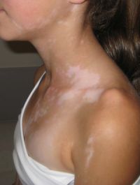
The defining feature is a flat, white or pale area of skin where melanocytes — the cells responsible for pigment — have been destroyed or stopped working. The edges of the patch are usually well-defined. Under a Wood’s lamp (ultraviolet light), depigmented areas glow blue-white because there is no melanin to absorb the UV.
The patches themselves are:
- Flat — not raised, scaly, or textured
- White to off-white — sometimes with a slightly pink or inflammatory border early on
- Asymptomatic in most cases — no itch, no pain, no soreness (occasional mild itching during an active phase is reported but not universal)
Hair growing from a depigmented patch may also turn white. This is called leukotrichia, and it can be an indicator of deeper melanocyte involvement — it tends to be harder to treat because the melanocyte reservoir in the hair follicle is also affected.
Where patches typically appear first
Vitiligo has a preference for certain areas, particularly where skin experiences friction or sun exposure:
- Face: around the eyes, mouth, and nose
- Hands and feet, especially the knuckles and fingertips
- Wrists and forearms
- Elbows and knees
- Around the genitals and inner thighs
- Axillae (armpits)
Mucous membranes — inside the mouth, nostrils, or around the eyes — can also be affected, though this is less common and more noticeable in people with darker surrounding skin.
The main types and what they mean for spread
Understanding the type matters because it affects both the prognosis and the treatment approach.
Non-segmental vitiligo (NSV) is by far the most common type, accounting for roughly 85–90% of cases. It is symmetric — patches tend to appear on both sides of the body in a mirrored pattern over time. It is associated with autoimmune activity and can spread unpredictably.
Segmental vitiligo (SV) affects one side or segment of the body only — often corresponding to a dermatome (the area served by a single nerve). It tends to progress quickly for 1–2 years and then stabilise. It is less associated with autoimmune activity and more resistant to phototherapy but may respond better to surgical options.
Focal vitiligo refers to one or a small number of isolated patches with no spread after 12+ months. Some cases stay focal indefinitely; others eventually become non-segmental.
Will it spread?
This is the question almost everyone asks first, and the honest answer is: it depends, and no one can reliably predict it.
In non-segmental vitiligo, spread is common but not inevitable. Some people have stable disease for years; others see new patches appear fairly quickly. Factors associated with more active spread include:
- A family history of vitiligo or other autoimmune conditions
- Recent emotional or physical stress
- Skin trauma — cuts, friction, sunburn (the Koebner phenomenon, where new patches appear at injury sites)
- Hormonal changes — pregnancy, thyroid disease
In segmental vitiligo, the pattern is typically faster early spread followed by stabilisation — often within 1–2 years of onset.
Active vs stable phases
Vitiligo moves between active phases (new patches forming, existing patches enlarging) and stable periods. During an active phase, patches may have a slightly reddish or inflamed border. Stable patches have sharp, clearly defined edges with no change over months.
Most treatment decisions — including when to start phototherapy or topical JAK inhibitors — are influenced by whether the disease is active or stable at the time.
What to monitor over time
If you have vitiligo, a practical approach is to:
- Photograph patches at consistent intervals (every 2–3 months) with the same lighting and distance
- Note any new patches that appear at sites of minor skin injury — this suggests the Koebner phenomenon is active
- Monitor for hair whitening within patches, which changes the treatment picture
- Get thyroid function checked if you haven’t — thyroid autoimmunity is the most common associated condition
Vitiligo itself causes no systemic harm, but its autoimmune mechanism means a higher statistical likelihood of other autoimmune conditions. A basic screen at diagnosis is worth doing once.
When to see a dermatologist
Sooner is better, for one specific reason: early-stage patches with no leukotrichia respond better to treatment. The window for repigmentation narrows once hair follicles in the patch are affected.
If you are newly diagnosed or noticing new patches, a dermatologist with experience in pigment disorders is the right starting point — both for confirming the diagnosis and for discussing whether treatment is warranted at this stage.
For an overview of what that first appointment typically looks like and what to expect from the diagnostic process: